Showing posts with label Eukaryotes. Show all posts

Gene mapping in eukaryotes


Mapping the genes in a correct order helps to know the location of the genes on the chromosomes. Various techniques achieved success in mapping the genes. Mapping techniques achieved success with eukaryotic organisms such as Drosophila and plants before humans. Gene mapping not only helps us in knowing the exact location of the gene but also helps in conducting various other experiments based on the gene location. Thomas Morgan worked with Drosophila strains and found out recombination mechanisms. An experiment sometimes gives an idea of other hidden strategies. For example, gene mapping studies revealed the mechanism of recombination. The studies revealed that the progeny obtained by crossing some strains of the eukaryotic organisms also showed phenotype differing from the parental phenotype.

Morgan’s experiment:
Thomas Morgan and his colleagues worked with Drosophila strains. They cultured Drosophila with a particular X-linked phenotype. The experimenters selected certain strains of fruit flies. The female flies had two X chromosomes with linked genes. The males had one X and one Y chromosome. The female flies had a phenotype of white eyes and miniature wings. The male flies were wild-type flies. The cross between these two types of flies gave rise to an F1 generation having wild-type females, and white-eyed, miniature winged male flies. Interbreeding of the F1 progeny gave different kinds of flies. There were total 2241 flies in the F2 generation. Out of these, total 900 flies had a non-parental phenotypic combination of white eyes and normal wings. Other types of non-parental strains included red-eyed, miniature winged flies. The non-parental ones are known as recombinants. The recombinants arise due to the crossing over between the homologous chromosomes. The theory involves two key concepts. The first one is the site of physical exchange. It is known as the chiasma. The second one involves the genetic recombination between the linked genes. It is known as crossing over. It also involves a reciprocal exchange of chromosome segments.


Image 2: Morgan's experiment

Stern’s Experiment:
Stern worked with X-linked gene loci in Drosophila. The experimenters conducted a cross between the wild-type bar eyed females and carnation type round-eyed males. The female flies had two X chromosomes. One of them additionally had a detached piece of X chromosome. The other X chromosome had an additional attachment of a piece of the Y chromosome. The chromosomes in the males flies had no extra pieces attached. The interbreeding of the F1 progeny gave rise to different types of flies. Four main types of progeny observed included carnation bar, red round, carnation round, and red bar eyed males and females respectively. The results of the experiment revealed the genetic recombination and exchange of identifiable segments. 

Barbara McClintock’s corn experiment:
The corn species selected for the experiment consisted of heterozygotes for the two genes on the 9th chromosome. One of the genes gave a phenotype of colored versus colorless. The other type of genes resulted in the phenotypes such as standard type starch with amylose and amylopectin versus waxy plants having the only amylopectin. The chromosomes had genes cWx giving normal phenotype. The homologs of the chromosomes having genes cWx had the genotype of Cwx. These homologs had a large double stained knob and a piece of 8th chromosome attached near the wx gene. It was a translocated segment. These features are known as the cytological markers. Hence, the corn experiments revealed the process of genetic recombination associated with the physical exchange between the parts of the homologous chromosomes.

Linkage studies using testcross:
A cross involving a normal individual with an individual who is homozygous recessive for all the genes is known as a testcross.
·        Two point test cross
Consider the autosomal recessive individuals. Suppose there involves a cross between the double heterozygotes with a genotype of a+b+/ a+b+ and double homozygous recessives with a genotype of ab/ab. The F1 generation revealed progeny with a wild-type a+b+/ab genotype. Upon conducting a testcross with double homozygous recessives, the progeny had 50% parental non-recombinants and 50% recombinant progeny. The formula for the recombination frequency involves (Number of recombinants/ Number of testcross progeny) x 100. The recombination frequency cannot exceed 50%.


Image 2: Two-point test cross

·        Three-point test cross:
Consider a cross between the triple heterozygotes with a genotype of a+b+c+/abc and triple homozygous recessives with a genotype of abc/abc. These crosses reveal the genetic recombination. Consider another example of flowering plants having three linked genes controlling the fruit phenotype. The recessive p allele gives a purple phenotype versus the wild-type yellow phenotype. The recessive r allele gives a round shape versus the wild-type elongated one. The recessive j allele gives juicy phenotype versus the wild-type dry fruit. The order of genes gets determined through a three-point test cross. Two parentals and six recombinants arise due to crossing over. The frequency of the double crossovers was found less than the frequency of the single crossovers.

Gene-centromere distance studies in Neurospora crassa:
The products of meiosis get a specialized arrangement depicting the four chromatids of each of the homologous pair of chromosomes. It usually reflects during the metaphase I. Neurospora consists of ordered tetrads. Meiotic and the mitotic divisions in the tetrads help in studying the process of recombination. It becomes easy to map the distance between the gene and the centromere using the ordered tetrads. The first division segregation tetrad consists of a parental type occupying half the ordered tetrad and another parental type in the other half of the tetrad. It occurs when there is no crossover. A single crossover between the gene and the centromere gives different types of tetrad segregation patterns (the second division segregation). The percentage of the second division tetrads divided by 2. It is known as the gene-centromere map distance. Tetrad analysis also helps in mapping two linked genes.

Mitotic recombination:
Crossing over is also known as genetic recombination between the linked genes or the reciprocal exchange of chromosome segments. It occurs during the mitosis as well as meiosis. The mitotic crossing over is also known as mitotic recombination. It leads to the production of the progeny cells having a combination of genes differing from the diploid parental cell entering the mitotic cycle. A classic example of the mitotic recombination includes fungus Aspergillus nidulans. It has a parasexual cycle of genetic systems. The genetic recombination in Aspergillus occurs through the processes other than regular alteration of meiosis and fertilization. The heterokaryon forms due to the mycelial fusion and the fusion of the two haploid nuclei. It gives rise to a diploid nucleus. The parasexual cycle also consists of mitotic crossing over within the diploid nucleus or haploidization of the diploid nuclei without meiosis. It becomes easy to calculate the gene order and the map distances.

Human gene mapping:
Physical mapping techniques help in mapping human genes. This technique mainly involves large genomes. It is not possible to set up a testcross for human genes since the human genome is vast. We obtain the recombination data from the pedigree analysis in humans. Gene mapping involves the use of gene markers and DNA markers.
                       
References:
[1] Genetics: Analysis of Genes and Genomes, Daniel L. Hartl, Elizabeth W. Jones
[2] Biology, Raven
[3] Biology, Pages 172-180, Neil A. Campbell, Jane B Reece
            

                                © Copyright, 2018 All Rights Reserved.

Eukaryotic cells undergo cyclical patterns of growth and division

The growth and developmental patterns observed in eukaryotic cells are different and complex as compared with prokaryotic cells. Also, the genetic material, the cellular compartmentalization, and mechanisms differ from that of prokaryotes. Eukaryotic cells are studied more than the prokaryotic cells because we humans are eukaryotes. The capacity of the cells to reproduce independently is tremendous. Hence, the study of the cell cycle helps to know exactly how a cell undergoes growth and division. The concept of two cells forming from a single cell gave rise to tissue and organ systems in the body. Thus it depends on how and when the cell undergoes a cyclical pattern. Every part of the cell has a specific role to play. Not only division but also the cells undergo differentiation. Thus there exist different types of cells such as immune cells, blood cells, sperm cells, and egg cells. The cells may be haploid or diploid containing one or two sets of chromosomes.

The genetic material is packed artistically in the cells. The chromosomes appear in different shapes when visualized under a microscope. We may describe a cell cycle as a process of cells in undergoing growth, replication of the genetic material and division. Before starting the discussion on the way cells undergo cyclical patterns, we must know the basic components of the cells. A eukaryotic cell structure consists of a nucleus and a cytoplasm. The nucleus is a house for the genetic material consists of DNA-protein interactive structures known as chromosomes. It is a place where DNA replication and transcription occurs. The extranuclear or outer part of the nucleus is known as the cytoplasm. It is a site for organelles and protein synthesis.

INFO BOX: Terminologies
1.     DNA: The genetic material in the nucleus. DNA is the blueprint of life.
2.     Chromosome: It is a linear structure composed of DNA-protein interaction.
3.    Chromatid: It is a subunit of the chromosome. The two chromatids constitute a chromosome. (Symmetrical halves running parallel to each other)
4.   Centromere: Chromatids are attached to each other through centromere. It is a constricted region.
5.     Centriole: It is a self-reproducing material involved in cellular processes.
6.  Nucleolus: It is RNA enriched spherical body associated with a chromosome segment.


Although a great variation exists in eukaryotic organisms and their cells, a typical eukaryotic somatic cell cycle duration is 24 hours. Four main stages of the cell cycle include the G1 phase, S phase, G2 phase, and M phase. The G1, S and G2 phases are clubbed and termed as interphase. The M phase or the mitotic phase is the division phase of the cell. The G1 phase is an initial period of growth or a pro synthetic gap with duration of ten hours. The S phase is the period of DNA synthesis. It lasts for nine hours. The G2 phase is a post-synthetic gap phase. It lasts for four hours. The duration of the mitotic phase is one hour. During the G1 phase, the chromosomes become thin and extended. The cell is responsive to growth signals. The G2 phase accompanies chromosome condensation. The cell cycle checkpoints regulate the transition from one phase to another. They monitor the integrity of the genome. The mitosis occurs in the somatic cells. Another process occurring in gametes is known as meiosis.

Mitosis:
The process of mitosis occurs in both haploid and diploid cells. Prophase, metaphase, anaphase, and telophase are stages of mitosis.
a.     Prophase: This is the initial stage of mitosis. It exhibits chromosome condensation and visibility. The spindle apparatus forms. A spindle is a set of tubulin fibers that move the eukaryotic chromosome during division. The spindle assembles outside the nucleus during the prophase. Early prophase, middle prophase, and late prophase are three subphases of prophase. In early prophase, the centrioles move apart. The chromosomes in early prophase reduce in their sizes. However, after some time they start getting visible. The nucleolus begins to disappear. The middle prophase involves the movement of the centrioles apart from each other. This phase is just a beginning of the mitotic spindle formation. The chromosomes are clearly visible in this phase. The late prophase involves the movement of centrioles towards the opposite sides. The spindle finally begins to form. The prophase chromosomes coil to produce a series of compact gyres. The kinetochore is a specialized protein that binds to the centromere. 
a.     Metaphase: It starts with the disappearance of the nuclear envelope. In this phase, the kinetochores are well attached to the centromeres so that the chromosomes align on the equator of the spindle. Hence their position is fixed on the equatorial plane. It is one plane halfway between the two spindle poles. The long axis of the chromosomes forms an angle of 900 to the spindle axis. A metaphase plate is an equatorial plane to which the chromosomes are aligned. During metaphase, the alignment of the chromosomes is perfectly on the spindle. The processed cells get arrested in the metaphase for visualizing under an electron microscope. Visualization under an electron microscope reveals a dense framework of proteins (scaffold) surrounded by an uncoiled DNA. Hence it reveals a double-stranded DNA.
b.    Anaphase: Here, the centromeres of the sister chromatids separate forming two daughter chromosomes. The two chromatids separate by moving under the action of the traction fibers towards the spindle pole. Along with the chromatids, the kinetochores also separate. Due to disjunction, the sister chromatids get converted to independent chromosomes and move towards the poles by shortening the microtubules. Thus, the chromosomes acquire specific shapes such as V, J or rod-shaped. The position of the centromere decides the shapes of the chromosomes. A submetacentric chromosome is J-shaped. A metacentric chromosome is V-shaped. Anaphase is very crucial for the cell due to centromere splitting. However, incorrect centromere splitting could be catastrophic for the cell.
c.      Telophase: The grouping of the daughter chromosomes occurs in such a way that the chromosomes align in two groups at the opposite ends of the cell. The chromosomes begin uncoiling and form elongated shapes. The spindle disappears. Reconstruction of the nuclear envelope begins. The nucleoli reappear. The nuclear division completes at this point. The cells get two nuclei.
d.    Cytokinesis: Division of the cytoplasm and the compartmentalization of the two nuclei occur into separate cells.


Image 1: Phases of Mitosis
Meiosis:
It is a process occurring in the gametes. It results in the doubling of the gametic chromosomes resulting in the zygotic chromosome number. The process of meiosis involves a single chromosomal duplication followed by two successive nuclear divisions. It occurs after one DNA replication cycle. The original diploid nucleus undergoes two successive divisions.  It consists of one haploid set of chromosomes from the father and the other from the mother. Meiotic division and differentiation lead to the formation of the gametes (the sperm and the egg). In the first meiotic division, the chromosome number gets reduced from diploid to haploid. The second meiotic division is equivalent to mitotic division. Four stages of meiosis I include prophase I, metaphase I, anaphase I and telophase I.
a.     Prophase I: It is somewhat similar to mitotic prophase. However, the difference lies in the behavior of the chromosome and the crossing over process. Five subphases of the prophase I include leptotene, zygotene, pachytene, diplotene, and diakinesis. The coiling of chromosomes occurs in the leptotene stage. Through this phase, the cells begin its commitment to meiosis. Zygonema is the early mid-prophase I. In this phase the shortening of the chromosomes takes place. Chromosomes align themselves roughly and are known as homologous chromosomes. They are similar to each other and synapse during meiosis. The homologous chromosomes retain their genes from the common ancestors. The process of synapsis leads to the formation of zipper-like structures along the length of the chromatids. The synaptonemal complex aligns the two homologs precisely. The telomeres initiate the process of synapsis. They get clustered on the nuclear envelope forming a bouquet like arrangements. Telomeres assist the chromosomes to undergo synapsis by moving them around. The mid-prophase or pachynema stage starts after the completion of the synapsis. Since the homologous chromosomes consist of four chromatids, they are known as bivalents or tetrad. Here, the crossing over or a physical exchange takes place. If there are genetic differences in the homologous chromosomes, crossing over results into formation of new genetic material. This crucial step leads to genetic variation and recombination. It just involves reciprocal exchanges. At the end of the pachynema, the synaptonemal complex disassembles and enters into diplonema stage. The homologous chromosomes begin to move apart. The crossing over, as the name suggests, begins to look like a cross. It is known as chiasmata where the homologous chromosomes associate tightly. In the diakinesis, the nuclear envelope breaks down with spindle assembly.
b. Metaphase I: There is a complete breakdown of the nuclear envelope with the alignment of bivalents on an equatorial plane. Spindle formation and microtubule attachment take place.
c.  Anaphase I: The chromosomes disjoin and migrate to the opposite poles. Hence maternally and paternally derived centromeres move towards each pole. The segregated sister chromatids remain attached at their respective centromeres.
d. Telophase I: Formation of the new nuclear envelope occurs. The cell proceeds for cytokinesis.
Meiosis II with prophase II, metaphase II, anaphase II and telophase II results in the formation of four haploid cells from two haploid cells of meiosis I. The results of meiosis II in males are known as spermatids. They are haploids giving rise to spermatozoa or sperms. The result of meiosis II in females is called ootid and a polar body.

References:
[1] Medical genetics, G.P. Pal
[2] Human Genetics, 3/e, Gangane
[3] Vogel and Motulsky's Human Genetics: Problems and Approaches, Friedrich Vogel, Gunter Vogel, Arno G. Motulsky
[4] Biology for the IB Diploma: Standard and Higher Level, Andrew Allott
[5] Principles of Medical Genetics, Thomas D. Gelehrter
© Copyright, 2018 All Rights Reserved.
Translation in eukaryotes

The process of translation occurs in the specialized structures known as ribosomes and requires a mRNA for initiating the process. Protein or a peptide is an arrangement of amino acids in a chain form. Hence, it is nothing but a linear arrangement of amino acids. The ribosome gets attached to the mRNA for initiating the process of translation. The process of translation is slightly different in eukaryotes. Unlike prokaryotic methionine, the eukaryotic methionine does not undergo modification. Also, the eukaryotic mRNAs do not have a Shine Dalgarno sequence. There is a different way of finding the AUG initiation codon.
Initiation:
A eukaryotic initiation factor known as Eif-4F gets involved in the process. A cap-binding protein (CBP) known as Eif-4E binds to the cap at 5’ end of mRNA. Scanning or finding an initiation codon across the mRNA in eukaryotes involves Met-tRNA, eIF proteins, and GTP. A short sequence known as a Kozak sequence consists of AUG codon. The process of scanning the initiation codon is known as a scanning model of initiation. The 40S ribosomal subunit binds to the AUG codon. Then the 60S subunit binds and displaces the eIFs. It produces an 80S initiation complex with the initiator tRNA bound to the mRNA in the P site of the ribosome. Since a mRNA molecule has a poly (A) tail binding protein (PABP) bound to a poly (A) tail, also binds to eIF-4G, thereby forming a loop close to the 5’ end. It is a sign of translation initiation in eukaryotes.
The initiation complex consists of mRNA, elongation factors (eIF4, eIF2, eIF3, and eIF5), 40S ribosomal subunit, and an initiator tRNA. The large subunit (the 60S) gets attached to a small subunit. The initiator tRNA forms a hydrogen bond with the AUG codon of mRNA.
The elongation and translocation steps are similar to that of prokaryotes. However, a small difference involves the number and the properties of the elongation factors and the sequence of the events. Both prokaryotes and eukaryotes employ a polyribosome or a polysome. Several ribosomes translate each mRNA simultaneously. A complex between mRNA and the ribosomes keeps translating simultaneously. It is known as a polyribosome or a polysome. Each ribosome in a polysome helps in producing a complete polypeptide chain.

Elongation:
The small ribosomal subunit (the 40S) moves one codon further on mRNA. The newly activated tRNA places itself in the large subunit. The new amino acid attaches to the previous amino acid under the influence of peptidyl transferase.

Termination:
The release factor binds with the ribosome on finding a stop codon. No new tRNA binds to the ribosome. Termination leads to detaching the polypeptide chain and releasing the factors.

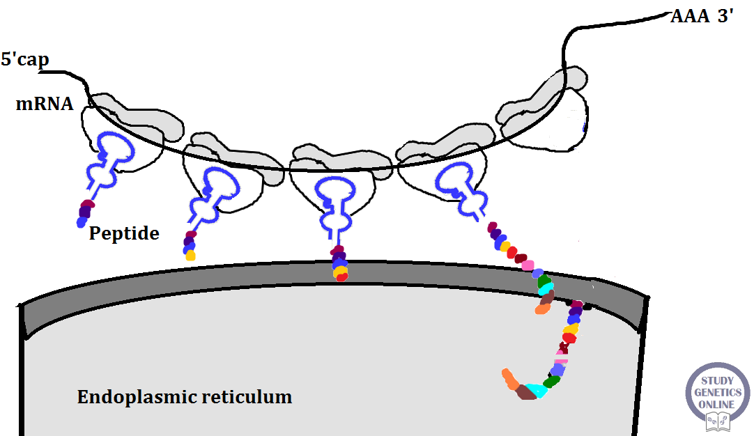
Image: Translation in the endoplasmic reticulum

Transfer RNA (tRNA):
It helps in transferring an amino acid to a growing polypeptide chain. A yeast tRNA contains 77 nucleotides. It is folded back upon itself and kept in a cloverleaf configuration. It involves a characteristic pairing of the bases G to C and A to U. Four important sites in a tRNA include a recognition site, an amino acid attachment site, codon recognition site, and ribosome recognition site. A recognition site is a specific base sequence recognizing a correct amino acid. The amino acid attachment site helps in the attachment of specific amino acid. A codon recognition site consists of three bases with a sequence complementary to a mRNA codon. The ribosomal recognition site helps the tRNA to recognize ribosome for the attachment. The tRNA attaches to the amino acid at the 3’ end. It contains a terminal adenylic acid followed by two cytidylic acids. Adjacent to the dihydrouridine loop lies a recognition site for a tRNA synthetase. The tRNA consists of certain rare bases formed after the transcription process. Examples include pseudo-uridylic acid (s), ribothymidylic acid (T), Dihydrouridylic acid (Ud), methyl guanylic acid (Gm), Dimethyl guanylic acid (Gd), Inosinic acid (I), and methyl inosinic acid (Im). A class of tRNA molecules known as Isoacceptor tRNA accepts the same amino acid but possesses different anti-codons. There are 2-4 Isoacceptor tRNA molecules in the higher organisms. They are specific to each amino acid.
Consider an example of a translation process occurring in the endoplasmic reticulum. The ribosome consists of the 50S and 30S subunits. Each ribosomal 50S subunit attaches to the membraneous component of the endoplasmic reticulum. The messenger RNA binds to the adjacent 30S subunit. The ribosomal 50S subunit consists of a cavity or a condensing site at the middle. The site gets occupied by a tRNA attached to a nascent polypeptide. Another site known as the entrance or the decoding site occupies both the subunits. It provides a site or a place for the aminoacyl tRNA which occupies the roof of the cavity. The uncharged tRNA gets removed after contributing its amino acid to the polypeptide. Suppose the sixth codon of the mRNA occupies a decoding site on the ribosome at the time T0. The CUG codon pairs with GAC anticodon of tRNA. After half a second, the mRNA advances to the next codon. It leads to an addition of a new amino acid to the nascent polypeptide chain.
References:
[1] Dictionary of Genetics, Himanshu Arora, 2007 edition.
[2] Crash Course: Cell Biology and Genetics E-Book, Matthew Stubbs, Narin Suleyman.
[3] Gene Regulation: A Eukaryotic Perspective, David S. Latchman


  © Copyright, 2018 All Rights Reserved.

Intricacies of Eukaryotic Transcription

The transcription process in eukaryotes involves a very complex process. It involves the synthesis of DNA followed by transcription. The complexity of the transcription process makes it important for us in knowing every detail. Studying transcription helps us in knowing more about the functionality of the molecules and processes known as protein synthesis. Unlike prokaryotic RNA polymerases, the eukaryotic polymerases involve three classes. After RNA synthesis, the molecule gets modified further. Hence, it is important to know the E. coli transcription process to clarify and study the same process in eukaryotes. Although similar to prokaryotes, eukaryotic transcription is different.

Eukaryotic RNA polymerases:
RNA polymerase-I is known to catalyze the synthesis of ribosomal RNA such as 28S, 18S, and 5.8S rRNA. RNA polymerase-II is known to synthesize messenger RNA (mRNA) and some small nuclear RNAs (snRNA). RNA polymerase-III synthesize transfer RNA (tRNA), 5SrRNA, and snRNA not synthesized by other polymerases. 

RNA synthesis:
The process of RNA synthesis is very complicated. The molecules involved in the process such as RNA polymerases also exhibit a complex nature. They have multiple subunits and encoded by several genes. There is very little information about these enzymes. Mainly RNA polymerase-II involves in transcribing the genes. The transcription process produces a precursor mRNA. It is modified to produce a mature and functional mRNA. Like prokaryotic cells, eukaryotic cells also consist of promoters. The promoter functions in eukaryotes involve more sophisticated technique as compared to prokaryotes. There are two parts of the promoter elements such as core promoters and promoter-proximal elements. The core promoters are known as cis-acting elements. These elements at 50 base pair upstream work well at or near the transcription site. They are necessary for starting the transcription process at the right site. Core promoter elements are short sequences or initiator (Inr) elements and TATA box elements. The Inr elements span the transcription site. The TATA box elements bind to RNA polymerase via TATA-binding proteins. These elements are analogous to Pribnow box in prokaryotes. The TATA box or Goldberg Hogness box consists of a seven nucleotide consensus sequence known as TATAAAA.

The core promoter also consists of general transcription factors (GTFs) required for initiation of transcription. The GTF labeling is specified based on the function. There are six types of GTFs such as TFIIA, TFIIB, TFIID, TFIIE, TFIIF, and TFIIH. TFIID binds to TBP of TATA box to form a complex. It is a binding site for other transcription factors such as TFIIA and TFIIB. Later on, TFIIF and RNA polymerase II bind and form a minimal transcription initiation complex. Later on, TFIIE and TFIIH bind to form complete transcription initiation complex. 

Core promoter
Normal function
Effect of mutation
Inr elements
Direct the initiation of transcription
Any mutation in the Inr elements makes the core promoter non-functional.
TATA Box
Helps in the binding of transcription factors and RNA polymerases to initiate transcription.
RNA polymerase cannot bind due to mutation. As a result, transcription does not occur.
Table 1: Promoters and their functions along with their effects after mutation. 

The promoter-proximal elements are present towards or near the place of attachment. In other words, they are upstream of the TATA box. There are two types of promoter-proximal elements such as CAAT box and GC box. The CAAT box consists of CAAT consensus sequence. The GC box consists of a consensus sequence GGGCGG. Both the promoter elements work in synergy. Mutation in these elements significantly affects the rate of transcription. A promoter function depends on both the core promoters and promoter-proximal elements. These elements control the time and way of gene expression. Transcription regulatory proteins help the promoter proximal elements to control the time and way of gene expression. These regulatory proteins are known as activators. The process of transcription becomes inefficient without the regulatory proteins. The promoter-proximal elements become incapable of regulating the transcription without activators.
Maximum efficiency in gene transcription involves another class of sequences known as enhancers. They are also known as cis-acting upstream or downstream elements. A peculiar specialty of an enhancer involves its maximum efficiency of transcription even from a distance. Many enhancers also determine spatial patterns of gene expression in higher eukaryotes.


Image 1: Initiation of transcription: It includes four main steps. (1) The initial commitment complex forms. The TATA box gets recruited along with the other proteins. (2) After the recruitment of TATA box and TBP, the other two transcription factors such as TFIIA and TFIIB join and initiate the transcription. (3) RNA polymerase-II along with TFIIH join the complex. (4) TFIIE and TFIIF bind and allow the completion of the transcription.

Initiation:
The transcription factors such as TFIID bind to TATA-binding protein on the TATA box. All other transcription factors also bind to this region along with the RNA polymerase II and form a pre-initiation complex. The RNA polymerase II starts synthesizing mRNA using only one strand of DNA double helix. The transcription begins at 5’ end and ends at 3’ end. Appropriate ribonucleotides get added to mRNA due to the activity of RNA polymerase II. The mRNA is a single-stranded molecule. All the sequences of structural genes including introns and exons get transcribed in mRNA. The mRNA is known as primary mRNA because it consists of introns that do not code for any protein. Hence they have the least importance in a functional mRNA. A process known as splicing removes the non-coding regions to give a fully functional RNA molecule.


Processing of pre-mRNA involves capping:
Immediately after synthesizing pre-mRNA, the process of capping occurs. Capping occurs after the RNA polymerase escapes the promoter. Hence, we must know two main processes such as promoter clearance and promoter escape. Promoter clearance involves moving ahead of the promoter sequences to begin RNA synthesis. Promoter escape means polymerase has completed its activity at the promoter site and now moves away from that region to proceed to cap. An extra guanosine group gets added to the extreme 5’ end of the RNA due to the activity of guanylyltransferase. The G- terminal gets methylated at the seventh nitrogen atom due to the activity of guanine methyl transferase. These two enzymes attach to the C-terminal domain of RNA polymerase II. 
Image 2: Steps involved in RNA synthesis and processing in eukaryotes.

Polyadenylation:
The process of polyadenylation accompanies the addition of the poly A tail to the 3’end. The tail is essential for exporting mRNA from the nucleus to the cytoplasm. It also protects it from exonuclease and stabilizes mRNA. Addition of a poly A tail to the 3’ end of RNA is a very complicated process in mammals. There is a poly A consensus sequence on mRNA known as AAUAAA sequence. CPSF also was known as Cleavage and Polyadenylation Specific factor and two other cleavage factor protein bind to and cleave the RNA. Then the enzyme poly-A polymerase adds a poly A tail to the 3’ end.

Elongation of eukaryotic mRNA:
Mammalian cells consist of 13 different elongation factors. The process of elongation starts when the RNA polymerase II has completed the promoter clearance or initiation process. The elongation factors exhibit various functions. The TFIIF, CSB, ELL, and Elongin are the factors suppressing the pausing of the RNA polymerase II. The pausing and stopping affect the activity of polymerases. Pausing arises due to the presence of a hairpin loop or intra-strand base pairs. Hence, these factors help the RNA polymerase II to skip such structures and move ahead. The TFII factor protects from the complete cessation of elongation. It is a chromatin modifier since it interacts with H2A and H2B.

mRNA with poly(A) tail
mRNA without Poly A tail
Helps in the termination process
Have two types of cleavage signals
A hairpin loop and a nine nucleotide consensus CAAGAAAGA
Table 2: Types of mRNA based on polyadenylation
Polyadenylation helps in terminating the process of transcription.

Termination
The termination of transcription is favored by changes in the CPSF-CTD interactions. The exact information on the mechanism of termination is not known.
The mRNA consisting of both protein-coding and non-coding region undergoes a process known as splicing. It is a post-transcription process that involves the removal of introns and ligation of exons to form a mature mRNA ready to depart from the nucleus.

References:
[1] Eukaryotic Transcription Factors, David Latchman, David S. Latchman
[2] Molecular Biology of the Cell, Bruce Alberts
[3] IGenetics, Peter Russell
[4] Gene Regulation: A Eukaryotic Perspective, David S. Latchman


© Copyright, 2018 All Rights Reserved

DNA Replication in Eukaryotes

The process of DNA replication involves duplication of DNA. It occurs by a semiconservative way in both prokaryotes and eukaryotes. There are specific chromosomal sequences found in the yeast Saccharomyces cerevisiae. Approximately 100 bp sequences are present in the yeast. These sequences can replicate autonomously. Such sequences are known as autonomously replicating sequences (ARS). There are three sequence elements known as A, B1, and B2. The origin recognition complex (ORC) is the initiator protein with a multi-subunit. The ORC binds to A and B1 in the yeast and recruits other proteins such as B2 DNA unwinding proteins. Between the B1 and B2 lies the replication origin. DNA replication occurs in the S phase of the cell cycle. The origin of replication is not used more than once in the cell cycle. There is a specific temporal ordering of initiation of replication. The replicator selection occurs in the G1 phase by assembling proteins on each of the replicator. They form pre-replicative complexes. Then the ORC recruits other proteins. Once the cell reaches the S phase of its cycle, the pre-replicative complexes get activated for initiating the replication. Cyclin-Dependent Kinase (CDK) enzyme activates the pre-RC to initiate the cell cycle. They also inhibit the formation of new pre-RCs. Except in the G1 stage, CDKs are present in all the phases of the cell cycle.


Image: DNA replication in eukaryotes

Eukaryotic replication enzymes:
Like prokaryotes, eukaryotes also require enzymes for replicating the DNA. There are more than 15 replicative enzymes in the eukaryotes. They are known as DNA polymerases. Pol α (Primase) is an enzyme that initiates new strands in replication by primase. It makes about ten nucleotides of an RNA which are further extended by Pol α. Formation of RNA-DNA primers occurs. Pol δ and Pol ε further extend these primers. Some of the functions of the replication enzymes of the eukaryotes are still not clear. There is no evidence about the role of specific enzymes in the synthesis of the leading and the lagging strand. Some other enzymes are involved in the replication of organelle DNA such as mitochondrial and chloroplast DNA. The mitochondrial DNA  replication follows a D-loop model. DNA polymerase γ is mainly involved in mitochondrial DNA replication. Another eukaryotic DNA polymerase is known as DNA Pol κ. The cohesion proteins hold the sister chromatids together until the anaphase of the nuclear division. The DNA Pol κ helps in the attachment of the cohesion proteins. DNA Pol δ works in synergy with an accessory protein known as proliferating cell nuclear antigen (PCNA), which is a functional equivalent of a β subunit of E.coli Polymerase III. PCNA holds the enzyme lightly to the template DNA.

The eukaryotic replication fork:
The DNA Pol α extends the initial RNA primer. Polymerase δ further replaces the Pol α. An enzyme is known as flap endonuclease (FEN1). It associates with the DNA Pol δ at the 3’ end of the Okazaki fragment. The primer from the 5’ end of the adjacent fragment gets degraded. However, the extreme 5’ end consists of a phosphate group which blocks the activity of the FEN 1. 
There are two models for studying the completion of the lagging strand in the eukaryotes.
a.     RNase H model: RNase H can remove the primer just to the last ribonucleotide. However, it cannot cleave the phosphodiester bond between the last ribonucleotide and the first deoxyribonucleotide. This ribonucleotide carries a 5’- monophosphate which can be removed by FEN-1. Thus the FEN-1 removes the last ribonucleotide and some of the DNA. The eukaryotic cell is capable of replicating DNA even in the absence of RNase H. Thus the discovery of the flap model came into existence.
b.    Flap model: The DNA Polymerase δ combines with the helicases for extending the Okazaki fragment. While the extension of the Okazaki fragment, the primer gets pushed aside. The extension of the exposed region occurs. A flap arises as a result of primer removal. FEN1 cuts this flap due to its endonuclease activity. FEN1 cleaves the phosphodiester bond at the branch point. Through the study of flap model, a few pieces of evidence revealed that the DNA Pol α may synthesize DNA in an error-prone way. DNA Pol α does not have 3’to 5’ proofreading activity. The helicase activity not only removes the primer but also eliminates the DNA that was originally synthesized by the DNA Pol α. On the other side, DNA Polymerase δ has a proofreading activity. Thus, it makes a highly accurate copy of a template.
There are certain problems in replicating the ends (telomeres) of the eukaryotic chromosomes. The parental DNA replicates and gives rise to two new DNA molecules. Each daughter DNA has an RNA primer at the telomere region. It is mainly at the 5’ end. The primase activity leads to the elimination of RNA primers. The primer removal leaves a single-strand stretch of DNA, thereby creating the gap at the 5’ end. DNA Polymerases cannot fill this gap. Thus the gaps may remain as they are. The leftover gaps may reduce the size of the chromosome after each replication. The process of aging is mainly due to the telomere shortening. The telomere replication has a specific mechanism. Eukaryotic chromosomes have tandem repeats at the telomeres.

Study of telomere replication in Tetrahymena:
The Tetrahymena is a protozoan. It has a repeated sequence of 5’-TTGGGG-3’ reading towards the DNA ends on the top strands. An enzyme known as telomerase contains protein and RNA subunits. The telomerase enzyme is responsible for maintaining the chromosome lengths. It adds repeats to the telomeres. Its RNA component has a sequence complementary to the telomere. The telomerase binds to the overhanging sequence and catalyzes the synthesis of three nucleotides TTG using its RNA component. The telomerase then slides towards the end of the chromosome. Its AAC region pairs with the newly synthesized TTG on the DNA. The rest of the telomere repeat is synthesized later on.

The Human DNA Replication:
The exact replication in human DNA is not extensively studied. However, few studies have described human DNA replication. The timing of the DNA replication varies in humans. Next to the lamin B2 gene on chromosome 19 is the origin of replication.

References:
[1] IGenetics, Peter Russell, second edition
[2] Eukaryotic DNA replication - Wikipedia

© Copyright, 2018  All Rights Reserved

Genomics and Proteomics for Cancer Research

The uncontrolled division of cells creates an abnormal environment in the body, leading to a condition known as cancer. It is the b...